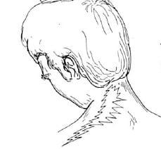
The most common cause of cervical nerve root compression (compressive neuropathology) is degenerative joint disease with stenosis and narrowing of the intervertebral foramen narrowing. The second most common cause is cervical disc herniation (1).
Cervical disc herniations with compressive neuropathology are commonly seen in asymptomatic populations (2, 3, 4). In a study published in 1996, 63% of asymptomatic men who were older than 40 years of age had protruding disks in the cervical spine (2). A study published in 1998 found demonstrable spinal cord compression secondary to cervical disc protrusion in 7.6% of asymptomatic subjects over the age of 50 years (3). However, it is rare for individuals to be asymptomatic with extruded disk herniations and spinal cord compression (4). This would mean that every day thousands of chiropractors are treating hundreds of thousands of patients with asymptomatic cervical spine disk herniations. It must be assumed that in a large percentage of these patients the chiropractor is performing high-velocity low-amplitude spinal adjusting at or adjacent to the level of the disk herniation.
Additionally, chiropractors routinely diagnose and treat cervical disk herniations on patients using a variety of approaches, procedures and techniques. Often, the chiropractor will utilize high-velocity low-amplitude spinal adjusting. Considering this, it is important to note that chiropractic spinal adjusting is extremely safe. The largest prospective study do date assessing the safety of chiropractic spinal manipulation to the cervical spine was published in Spine, October 2007, and titled (5):
Safety of Chiropractic Manipulation of the Cervical Spine
A Prospective National Survey
This study assessed the risk of serious and relatively minor adverse events following chiropractic manipulation of the cervical spine by a sample of United Kingdom chiropractors. The authors studied treatment outcomes from 19,722 patients from 377 chiropractors that involved 50,276 cervical spine manipulations. Manipulation was defined as the application of a high-velocity/low-amplitude or mechanically assisted thrust to the cervical spine. A serious adverse event was defined as referred to hospital and/or severe onset/worsening of symptoms immediately after treatment and/or resulted in persistent or significant disability/incapacity, and minor adverse events reported by patients as a worsening of presenting symptoms or onset of new symptoms, were recorded immediately, and up to 7 days, after treatment. The authors concluded:
“Safety of treatment interventions is best established with prospective surveys, and this study is unique in that it is the only prospective survey on such a large scale specifically estimating serious adverse events following cervical spine manipulation.”
“There were no reports of serious adverse events.”
“Although minor side effects following cervical spine manipulation were relatively common, the risk of a serious adverse event, immediately or up to 7 days after treatment, was low to very low.”
“No significant adverse event was reported by the chiropractors using the definition criteria.”
“The risk rates described in this study compare favorably to those linked to drugs routinely prescribed for musculoskeletal conditions in general practice.”
“The risks reported here are also lower than those reported for acupuncture, which were described as a very safe intervention in the hands of a competent practitioner.”
“Although minor side effects were found to be relatively common, the risk of a serious adverse event, immediately and up to 7 days after treatment, was estimated to be low to very low in these consultations.”
“On this basis, this survey provides evidence that cervical spine manipulation is a relatively safe procedure when administered by registered United Kingdom chiropractors.”
Epidemiological studies show that the most common cervical disk herniations are C5-C6 (affecting the C6 nerve root) and the C6-C7 (affecting the C7 nerve root) levels (6). [Remember, since the C1 nerve root exits between the occiput and the atlas, there are 8 cervical nerve roots. This is why the C6 nerve roots exits between C5-C6 vertebrae, and the C7 nerve root exits between C6-C7 vertebrae].
•••••••••
Diagnosing Cervical Disk Herniation with Compressive Nerve Root Pathology Clinical Signs and Symptoms Diagnostic Imaging
Acknowledging that cervical disk herniation can be asymptomatic, even in the presence of apparent imaging of compressive nerve root neuropathology, classic clinical presentation would include combinations of the followings:
- Neck pain.
- Arm pain, usually extending below the elbow.
- Alterations of superficial sensation in a dermatomal pattern. This can be either hypoesthesia or hyper I often find that in acute situations there is hyperesthesia. In the subacute and/or chronic situation, there is hypoesthesia. According to Stanley Hoppenfeld, MD [Associate Clinical Professor of Orthopaedic Surgery Albert Einstein College of Medicine, New York] (7):
- C5 nerve root would involve the lateral arm.
- C6 nerve root would involve the lateral forearm, thumb, and 2nd
- C7 nerve root would involve the 3rd (middle) phalange.
- C8 nerve root would involve the 4th (ring) and 5th (little) phalanges.
- Reduced segmental sensory-motor (deep tendon) reflex (7):
- C5 nerve root: biceps tendon
- C6 nerve root: brachioradialis tendon
- C7 nerve root: triceps tendon
- C8 nerve root: none
- Myotomal muscle weakness is the weakness of a muscle associated primarily with a single nerve root. Again, as established by Stanley Hoppenfeld, MD, (7):
- C5 nerve root: the deltoid muscle; arm abduction
- C5 nerve root: the biceps muscle, elbow flexion
- C6 nerve root: wrist extension
- C7 nerve root: triceps muscle, elbow extension
- C7 nerve root: wrist flexion, finger extension
- C8 nerve root: finger flexion
| Shoulder Abduction | C5 | Deltoid |
| Elbow Flexion | C5 | Biceps |
| Elbow Extension | C7 | Triceps |
| Wrist Extension | C6 | Wrist Extensors |
| Wrist Flexion | C7 | Wrist Flexors |
| Finger Extension | C7 | Finger Extensors |
| Finger Flexion | C8 | Finger Flexors |
- Positive extremity pain exacerbation with cervical spine compression:
- Neutral position
- Lateral flexed position
- Spurling’s test (lateral flexion with ipsilateral rotation [from (13)]):

- Positive extremity pain with shoulder depression (shoulder depression test) (9).
- Pain relief with cervical spine distraction.
- Increased arm pain with the upper limb tension test (arm abducted 90°, elbow flexed 90°, fingers and wrist extended; followed by slowly extending the elbow).
- Aggravation of neck and/or arm pain with an increase in intrathecal pressure: Valsalva test, sneezing, coughing, and straining at stool.
- Diagnostic imaging findings, typically an MRI, showing compressive neuropathology at the level consistent with the other motor and sensory findings.
•••••••••
Studies on the Non-Pharmacological, Non-Surgical Management of Cervical Disk Herniation
•••••••••
In 1996, physicians (and brothers) Joel and Jeffery Saal, published a study in the journal Spine and titled (10):
Non-operative Management of Herniated Cervical
Intervertebral Disc With Radiculopathy
These authors note that frequently, patients with cervical disc herniation and neurologic loss or radiculopathy, are treated surgically if they have symptoms that persists after rest or minimal intervention. They state:
“For non-validated reasons, cervical disc extrusions have been frequently considered a definite indication for surgery.”
Consequently, they evaluated the clinical outcomes in 26 patients with cervical herniated nucleus pulposus and radiculopathy who were treated with an aggressive physical rehabilitation program. These patients were followed for longer than 1 year.
Inclusion criteria for this study included a focal cervical disc protrusion of less than 4 mm identified on magnetic resonance imaging and a major complaint of extremity pain compatible with cervical radiculopathy. Exclusion criteria included severe central canal stenosis and/or symptomatic cervical myelopathy.
All patients had a predominance of radicular upper extremity pain as their chief complaint. All patients had a magnetic resonance imaging (MRI) showing a cervical herniated nucleus pulposus that extended at least 4 mm from the margin of the parent disc space. Twenty of twenty-six (20/26) patients had extruded cervical discs. Six of the twenty-six (6/26) patients had contained disc herniations.
The rehabilitation program management consisted of traction, specific physical therapeutic exercise, and patient education. All patients were treated with ice, relative rest, a hard cervical collar worn for up to 2 weeks in a position to maximize arm pain reduction (all patients), manual and mechanical traction in physical therapy, followed by home cervical traction, and progressive strengthening exercises of the shoulder girdle and chest with training in postural control and body mechanics training. The duration of this portion of the program was 3 months, at which time the patient was discharged to an independent exercise program. The non-operative treatment in the patients in the present study averaged 9 months. Forceful joint manipulation was not used.
Twenty-four patients (92%) were successfully treated without surgery.
Twenty patients achieved a good or excellent outcome, and of these 19 had disc extrusions. Two patients underwent cervical spine surgery. Twenty-one patients returned to the same job. High patient satisfaction with non-operative care was achieved on outcome analysis.
The authors concluded:
“Many cervical disc herniations can be successfully managed with aggressive nonsurgical treatment.”
“A small percentage of patients with cervical herniated nucleus pulposus do require surgery for radiculopathy. However, the majority can be treated successfully with a carefully applied and progressive non-operative program.”
“The presence of radicular neurologic loss or nuclear extrusion should not be used solely as the criterion for surgical intervention.”
A couple of side observations by these authors include noting that inflammation of neural elements appears to play an important role in radiculopathy. Additionally, they state:
“Reabsorption of extruded disc material itself probably occurs in the cervical HNP as it does in the lumbar disc HNP.” Because of this reabsorption, “an extruded disc actually may have a more favorable non-operative prognosis than contained disc pathology.”
The most important point made by this article is that herniated cervical discs, even extrusion of the cervical disc, with radiculopathy (motor and sensory signs), can be successfully conservatively managed by a regime that consists primarily of exercise, traction, and mobilization. Another important point is that herniated cervical discs, even extrusion of the cervical disc, with radiculopathy (motor and sensory signs), rarely require surgery, even if they have significant extremity weakness or severe pain longer than 8 weeks. Most importantly, herniated cervical discs, even extrusion of the cervical disc, with radiculopathy (motor and sensory signs), should always be conservatively managed before surgery is warranted.
•••••••••
In 1999, physician Brian Nelson and colleagues published a study in the journal Archives of Physical Medicine and Rehabilitation titled (11):
Can spinal surgery be prevented by aggressive strengthening exercises?
A prospective study of cervical and lumbar patients
All 46 of the patients in this study had been recommended for spinal surgery. The objective of this study was to assess if an aggressive strengthening program could help these patients to avoid the surgery. Nine of these patients were diagnosed with “cervical disc syndrome” [distinct from degenerative cervical disc disease].
The aggressive strengthening program was 10 weeks in duration. It consisted of intensive, progressive resistance exercise of the isolated lumbar or cervical spine. The exercises were continued until fatigue failure, and patients were encouraged to work through their pain. “An important point is that the training was quite vigorous and did not stop because of pain exacerbation.” Patients were encouraged to be vigorous. They were taught, “hurt does not necessarily mean harm.” The average follow-up was at 16 months following discharge.
None of the cervical patients underwent surgery in the follow-up period.
“The study is valuable because it shows that a large number of surgical candidates at a private practice clinic can avoid surgery over an extended period. Further, there were no significant complications or negative consequences associated with delaying surgery while patients participated in an aggressive strengthening program. Occasional exacerbations occurred, but these were self limited and did not prevent rehabilitation from continuing.”
“Surgical candidates are often considered more ‘fragile’ than non-surgical patients and are more often guided toward inactivity to protect the spine. Many have been told to remain inactive based on MRI scans. They develop a keen sense of fear when it comes to spinal motion. Spinal pain patients become expert at substituting pelvic or thoracic movement for lumbar or cervical motion, respectively. In this way they protect the injured body part from meaningful exercise.”
“Substitution protects the lumbar or cervical spine from normal movement. Without motion the disc deteriorates, disc pH decreases, joints stiffen, ligaments shorten, bone density decreases, and muscles become deconditioned. Recent evidence suggests that a damaged disc becomes more acidic and that reduced pH is a mediator of spinal pain. The adult disc is an avascular structure that depends on diffusion for its nutrition. Diffusion is facilitated by a pumping action through spinal motion. Lack of motion, however, hinders diffusion. In the aggressive strengthening program in this study, patients were not allowed to substitute. The cervical and/or lumbar spine was isolated in such a way that substitution was impossible. Exercise therefore facilitated fluid exchange in the disk, which may account for the subjective improvement.”
“The significance of this study is that many patients were spared surgery during the study period even though surgery had been recommended. The findings show that a percentage of spinal patients can avoid surgery by completing an aggressive strengthening program and that even patients recommended for spinal surgery can tolerate intensive, specific exercise.”
Although this study did not involve joint adjusting, there emerged an important physiological concept for chiropractors who are managing disc herniation patients:
The pain of disc herniation and radiculopathy may be more due to changes in chemistry (inflammation, acidity) than to the actual compression. Understandably, such patients reduce the motion of the injured joints to avoid increased pain perception. However, reduced motion compromises tissue integrity and enhances the pathology. Without motion, the disc further deteriorates because of lack of nutrition though diffusion mechanisms, becoming more acidic and more painful.
Theoretically, any treatment that restores motion to the motor segment would improve diffusion, improve disc chemistry, and reduce pain while improving function. In this study, intense isolated spinal exercises were able to accomplish this goal. Is there another approach to restoring spinal biomechanical motion that is both effective and safe?
•••••••••
Cynthia Peterson, DC, and colleagues, from the University of
Zurich, Switzerland, published an important study last month (October 2013) pertaining to the use of chiropractic High-Velocity Low-Amplitude Spinal Manipulation in patients with cervical disc herniations. Their article was published in the Journal of Manipulative and Therapeutics, and titled (12):
Outcomes From Magnetic Resonance Imaging:
Confirmed Symptomatic Cervical Disk Herniation Patients Treated With High-Velocity, Low-Amplitude Spinal Manipulation Therapy:
A Prospective Cohort Study With 3-Month Follow-Up
Dr. Peterson is a Professor, Department of Chiropractic Medicine, Faculty of Medicine, Orthopedic University Hospital Balgrist, University of
Zurich, Switzerland. The purpose of her study was to investigate outcomes of patients with cervical radiculopathy from cervical disk herniation (CDH) who were treated with spinal manipulative therapy.
This study used 50 patients with a mean age of 44 years. Chiropractic treatment frequency was 3 to 5 times per week for the first 2 to 4 weeks and then 1 to 3 times per week thereafter until the patient was asymptomatic. Patients were evaluated at baseline, 2-weeks, 1-month, and 3-months.
The patients in this study had neck pain and moderate to severe arm pain in a dermatomal pattern with sensory, motor, or reflex changes corresponding to the involved nerve root. Patients also had at least one of the following positive orthopedic tests for cervical radiculopathy:
- Positive upper limb tension test
- Positive cervical distraction test
- Positive Spurling’s test
- Positive cervical rotation test at less than 60°
Finally, all of these patients had a magnetic resonance imaging–proven cervical disc herniation at the corresponding spinal segment.
The measurement outcomes used to assess these patients included:
- The Numeric rating scales (NRS) for pain where 0 is no pain and 10 is the worst pain imaginable for both the neck and the arm pain separately.
- The Neck Disability Index (NDI).
The chiropractic spinal adjustment given to these patients was described as follows:
“High-velocity, low-amplitude spinal manipulations were administered by experienced doctors of chiropractic.”
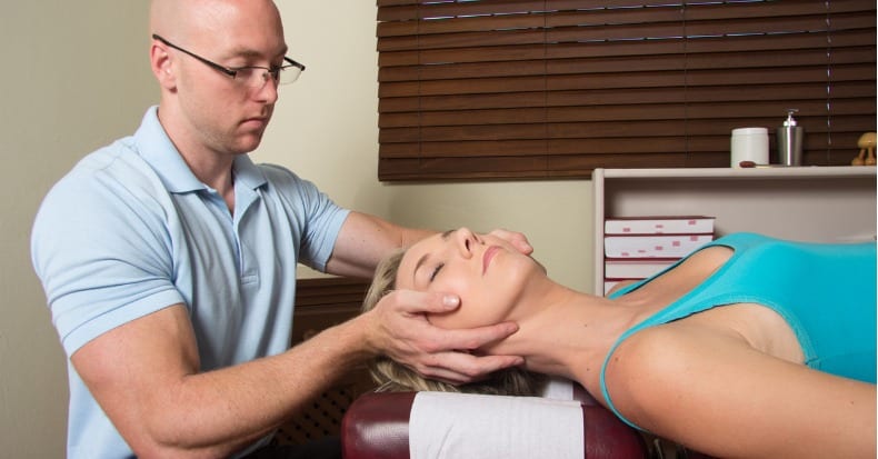
“The treatment procedure was a standardized, single, high-velocity, low-amplitude cervical manipulation with rotation to the opposite side and lateral flexion to the same side of the affected arm.”
“The chiropractor stood on the affected side of the supine patient’s neck, with an index contact on the articular pillar of the most symptomatic vertebral motion segment on the side of the patient’s complaint and at the spinal level clinically assessed to correspond with the MRI findings.”
“Rotation to the opposite and lateral flexion to the ipsilateral side was used to take out skin and joint slack.”
“Once the patient was positioned, a high-velocity, low-amplitude thrust was applied, with the goal of moving the affected segment and producing an audible release.” An audible release was achieved in most case.
“In the rare case where an audible release did not occur during the procedure, the chiropractor might repeat the manipulation up to 2 additional times.”
“When a patient reported bilateral neck and/or arm pain (extremely rare), the procedure could be reproduced on the opposite side as well.”
The measured outcomes for these patients were noted as follows:
“By 2 weeks after the first treatment, 55.3% of all patients reported that they were significantly improved and none reported being worse.”
“At 1 month, 68.9% were significantly improved.”
By 3 months 85.7% were significantly improved with no patients being worse.
“Statistically significant decreases in neck pain, arm pain, and NDI scores were noted at 1 and 3 months compared with baseline scores.”
For the subacute/chronic patients, the mean duration of symptoms was 299 days. At 3 months, 76.2% of these patients reported clinically relevant improvement with no patients reporting that they were worse.These authors make these following comments:
“Most patients in this study, including subacute/chronic patients, with symptomatic magnetic resonance imaging–confirmed cervical disc herniation treated with spinal manipulative therapy, reported significant improvement with no adverse events.”
“Most patients in this study with MRI-proven symptomatic cervical disc herniations who were treated with high-velocity, low-amplitude spinal manipulation reported clinically significant improvement at all time points, particularly at 3 months.”
“It is important to point out that even the subacute/chronic patients in this study with symptoms lasting longer than 4 weeks (mean duration, 298.73 days) reported high levels of clinically significant improvement. This is clinically important as the chronic patients are the ones who are usually the most costly in terms of health care use and quality-of-life disruption.”
Although subacute/chronic patients responded extremely well in this study, overall acute patients showed faster and greater improvement.
•••••••••
Cervical disc herniations are commonplace in asymptomatic populations. All chiropractors occasionally see patients with cervical disc herniation and clinically correlated compressive neuropathology. Effective and safe management of these patients includes traction, mobilization, stabilization exercises, aggressive strengthening exercise program, and spinal adjusting at the level and side of the herniation. These studies suggest that the patient’s pain and disability are linked to local chemical changes, which themselves are linked to fear avoidance and biomechanical reductions of motion. Consequently, clinical improvement is dependent upon the restoration of movement, and specific chiropractic adjustments are safe and effective in this regard.
References
- Radhakrishnan K; Litchy WJ; O’Fallon WM; Kurland LT; Kurland LT; Epidemiology of cervical radiculopathy; A population-based study from Rochester, Minnesota, 1976 through 1990; Brain 1994;117; pp. 325-35.
- Healy JF, Healy BB, Wong WHM, Olson EM; Cervical and lumbar MRI in asymptomatic older male lifelong athletes: frequency of degenerative findings; Journal of Computer Assisted Tomography;1996; Vol. 20; pp. 107-112.
- Matsumoto M, Fujimura Y, Suzuki N, et al; MRI of cervical intervertebral discs in asymptomatic subjects; Journal of Bone and Joint Surgery (British); 1998; Vol. 80-B; pp. 19-24.
- Ernst CW, Stadnik TW, Peeters E, Breucq C, Osteaux MJC; Prevalence of annular tears and disc herniations on MR images of the cervical spine in symptom free volunteers; European Journal of Radiology; 2005; Vol. 55; pp. 409-414.
- Thiel HW, Bolton JE, Docherty S, Portlock JC. Safety of chiropractic manipulation of the cervical spine. A prospective national survey. Spine; 2007; Vol. 32; pp. 2375-2378.
- Radhakrishnan K, Litchy WJ, O’Fallon WM, Kurland LT, Kurland LT. Epidemiology of cervical radiculopathy. A population-based study from Rochester, Minnesota, 1976 through 1990. Brain; 1994; Vol. 117; pp. 325-335.
- Hoppenfeld S; Orthopaedic Neurology: A Diagnostic Guide to Neurologic Levels; Lippincott, 1977.
- White AA, Panjabi MM; Clinical Biomechanics of the Spine; Lippincott; 1990.
- Jackson R; The Cervical Syndrome; Thomas; 1978.
- Saal, Joel S. MD; Saal, Jeffrey A. MD; Yurth, Elizabeth F. MD; Nonoperative Management of Herniated Cervical Intervertebral Disc With Radiculopathy; Spine; Volume 21(16) August 15, 1996, pp. 1877-1883.
- Brian W Nelson, David M Carpenter, Thomas E Dreisinger, Michelle Mitchell, Charles E Kelly, Joseph A Wegner; Can spinal surgery be prevented by aggressive strengthening exercises? A prospective study of cervical and lumbar patients; Archives of Physical Medicine and Rehabilitation; January 1, 1999; Vol. 80; No. 1; pp. 20-25.
- Peterson CK; Schmid C; Leemann S; Anklin B; Humphreys BK; Outcomes From Magnetic Resonance Imaging: Confirmed Symptomatic Cervical Disk Herniation Patients Treated With High-Velocity, Low-Amplitude Spinal Manipulation Therapy: A Prospective Cohort Study With 3-Month Follow-Up; Journal of Manipulative and Therapeutics; October 2013; Vol. 36; pp. 461-467.
- White AA, Panjabi MM; Clinical Biomechanics of the Spine; 2nd Edition; Lippincott; 1990; p. 410.
