When a whiplash event occurs—such as a motor vehicle collision—the rapid back and forth motion of the head can injure the bones, muscles, ligaments, joints, and nerves (including the spinal cord) in the neck leading to the collection of symptoms known as whiplash associated disorders (WAD). While about 50% of those injured can expect a rapid recovery, the other half has longer term complaints, which may include neck pain, headache, widespread sensory hypersensitivity, psychological distress, and changes in motor performance, cognitive function, and muscle composition. One highly frustrating aspect of WAD injuries is the lack of objective tests to make a firm diagnosis. However, a recent study may have identified a new tool in diagnosing injury to the spinal cord, which may help identify patients at risk for ongoing issues.
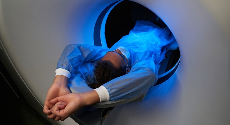
Routine x-ray is not sensitive enough to pick up cord-related injury in the absence of obvious fracture or dislocation. Though there are some clinical exam tests that can help identify minor spinal cord injury such as heightened reflexes and abnormal brain/cord-related superficial reflex signs, a new study describes an advanced MRI method, magnetization transfer imaging (MTI), that can quantify a discrete cord injury. Of the seventy-six WAD patients in the study, thirty reported complete recovery at the one-year point, thirty-two continued to experience mild symptoms, and fourteen had severe ongoing symptoms. A review of each patient’s initial neck disability index (NDI) questionnaire revealed that the patients who did not recover had higher pain and disability scores early on. However, there was no difference in initial NDI scores in the mild/severe one-year symptom groups.
The researchers then used MTI to look for signs of injury in different tracts of the spinal cord and found differences in the areas that transfer motor and sensory information between all three groups. Investigators add that abnormal findings were more common among the female participants, as women are nearly two times more likely to develop chronic WAD. It’s suspected this may be due to anatomical differences between male and female necks with respect to muscle mass and length.
Chiropractors routinely utilize questionnaires such as the NDI and clinical neurological reflex tests to track a patient’s course of care and response to treatment. When signs and symptoms persist, advanced imaging such as MRI can shed additional light as to why those patients have not recovered. Until MTI becomes more available, reliance on the current diagnostic tools (questionnaires and clinical exam) will continue to be the standard.
The good news is that there are MANY clinical studies that support chiropractic’s multi-modal treatment approach in the management of WA that includes manual therapies (manipulation, mobilization, soft tissue release techniques, etc.), physical modalities that promote healing, exercise training for soft tissue strengthening, and neurological recovery (balance, visual disturbances, light/noise sensitivity, etc.). As technologies continue to advance and become more readily available, healthcare providers will be able to better identify physical reasons for ongoing pain and provide appropriate interventions in attempt to prevent chronic, long-term complaints.
